
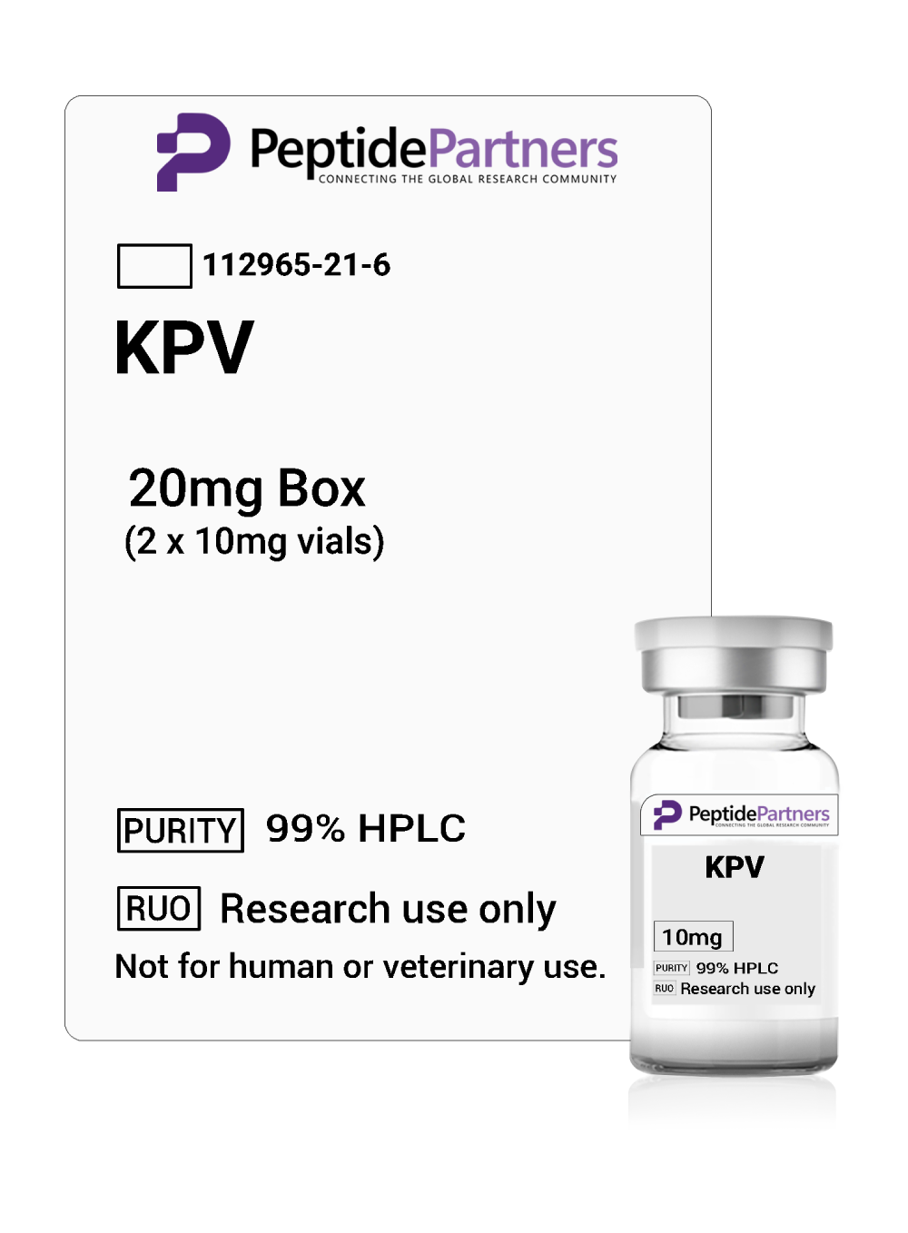
KPV (10mg vials)
Price range: $88.00 through $320.00
• Purity: 99.60% (multi-vial, independently tested)
• Format: 10mg vials (3 mL capacity)
• Box Options: 20mg, 50mg, 100mg combinations
• Testing Status: Endotoxin, heavy metals & sterility screening PASSED
• Cost Efficiency: $3.20 – $4.40 per milligram
KPV (lysine-proline-valine) is a tripeptide fragment derived from alpha-melanocyte-stimulating hormone (α-MSH) used in preclinical research to study melanocortin receptor pharmacology, inflammatory signaling pathways, and peptide transporter mechanisms. This research-grade material supports in-vitro experimentation focused on NF-κB pathway modulation, cellular uptake kinetics, and anti-inflammatory compound characterization.
- Independent certificate details, manufacturer IDs, and batch IDs should be reviewed before purchase when available.
- Order cancellations for full refunds are available before shipment; shipped orders cannot be cancelled or refunded.
- For damaged or incorrect goods, photograph the outer packaging, inner packaging, and product labels, then email support within 48 hours of carrier delivery.
Buy KPV – Peptide Partners
Product Overview & Specifications
KPV (lysine-proline-valine) is a tripeptide fragment derived from alpha-melanocyte-stimulating hormone (α-MSH) investigated for its role in inflammatory signaling modulation and melanocortin receptor interactions. Peptide Partners supplies this research-grade material for in-vitro testing, laboratory experimentation, receptor pharmacology, cellular uptake studies, and preclinical inflammation research.
Published studies have examined KPV using intestinal epithelial cell lines, keratinocyte cultures, bronchial epithelial cells, peptide transporter assays, NF-κB reporter systems, and three-dimensional skin models. Research areas include PepT1-mediated cellular uptake, NF-κB pathway inhibition, MAPK signaling modulation, oxidative stress responses, nuclear import mechanisms, and melanocortin receptor subtype selectivity.
The product name supplied for this listing is “KPV.” Researchers should verify the exact identity, sequence, formulation, and batch-specific certificate of analysis before beginning any experiment. Findings from published KPV studies should not be assumed to apply to every commercial batch or formulation.
Product Specifications
| Specification | Details |
|---|---|
| Product name | KPV (Lysine-Proline-Valine) |
| Compound type | Tripeptide (α-MSH fragment) |
| Primary molecular targets | Peptide transporter 1 (PepT1), NF-κB pathway, MAPK signaling, melanocortin receptors |
| Primary research areas | Inflammatory signaling modulation, cellular uptake kinetics, NF-κB pathway inhibition, oxidative stress responses, and melanocortin receptor pharmacology |
| Product format | 10 mg vials |
| Vial size | 10 mg |
| Vial capacity | 3 mL |
| Available box combinations | 20 mg, 50 mg, and 100 mg |
| Multi-vial purity | 99.60% |
| Independent testing | Yes |
| Endotoxin screening | Passed |
| Heavy-metals screening | Passed |
| Sterility screening | Passed |
| Manufacturer ID | WF03 |
| Batch ID | KV202606 |
| Cost per milligram | $3.20–$4.40 |
The stated 99.60% purity is based on multi-vial testing. Researchers should review the batch-specific certificate of analysis before beginning any experiment.
Primary Research Studies & Findings
PepT1-Mediated Tripeptide KPV Uptake Reduces Intestinal Inflammation
Authors: Guillaume Dalmasso, Laetitia Charrier-Hisamuddin, Hang Thi Thu Nguyen, Yutao Yan, Shanthi Sitaraman, and Didier Merlin
Publication: Gastroenterology, 2008;134(1):166–178
PMCID: PMC2431115
Reference: View publication
This study investigated the anti-inflammatory mechanisms of KPV in human intestinal epithelial cells (Caco2-BBE and HT29-Cl.19A) and human T cells (Jurkat) stimulated with pro-inflammatory cytokines. Nanomolar concentrations of KPV inhibited the activation of nuclear factor-kappa B (NF-κB) and mitogen-activated protein (MAP) kinase inflammatory signaling pathways and reduced pro-inflammatory cytokine secretion.
Uptake experiments using radiolabeled tritiated KPV ([3H]KPV) determined kinetic characteristics of KPV cellular uptake. The study found that KPV acts via human peptide transporter 1 (hPepT1) expressed in both immune and intestinal epithelial cells. KPV anti-inflammatory effects were assessed using NF-κB luciferase gene reporter assays, western blot analysis for signaling proteins, real-time RT-PCR for gene expression, and ELISA for cytokine quantification.
The researchers demonstrated that KPV is transported into cells by PepT1 and exerts anti-inflammatory effects through inhibition of pro-inflammatory signaling pathways at concentrations as low as the nanomolar range. The findings suggest KPV’s potential as a research tool for studying intestinal inflammation and peptide transporter-mediated drug delivery systems.
Plain-English Research Summary
This research showed that KPV, a tiny three-amino-acid peptide, can reduce inflammation in intestinal and immune cells grown in the laboratory. The peptide enters cells through a specific transporter protein called PepT1, which normally helps absorb nutrients from food. Once inside the cells, even very small amounts of KPV block the activation of key inflammatory signaling pathways, particularly NF-κB, which is like a master switch for inflammation. This prevents the cells from producing inflammatory chemicals called cytokines.
Lysine-Proline-Valine Peptide Mitigates Fine Dust-Induced Keratinocyte Apoptosis and Inflammation by Regulating Oxidative Stress and Modulating the MAPK/NF-κB Pathway
Authors: Junghee Sung, Seo-Young Ju, SeungHyun Park, Won-Kyo Jung, Jae-Young Je, and Sei-Jung Lee
Publication: Tissue and Cell, Volume 95, August 2025, 102837
DOI: 10.1016/j.tice.2025.102837
Reference: View publication
This study investigated the protective effects of KPV against oxidative damage and inflammation induced by fine particulate matter (PM10) in human HaCaT keratinocytes using in vitro cell culture methods. HaCaT cells were exposed to PM10 at different concentrations (0–200 μg/mL) for 24 hours, with significant cytotoxic effects observed at concentrations between 100 and 200 μg/mL.
Treatment with 50 μg/mL of KPV restored cell viability and reduced interleukin-1 beta (IL-1β) secretion that had been disrupted by PM10 exposure. KPV inhibited reactive oxygen species (ROS) production, which is responsible for activating extracellular signal-regulated kinase (ERK) and p38 mitogen-activated protein kinase (MAPK). KPV decreased the expression of apoptosis-related proteins including Bax, Bcl-2, and cleaved caspase-3, as well as IL-1β, through suppression of the redox-sensitive transcription factor NF-κB in PM10-treated HaCaT cells.
KPV effectively blocked ROS-mediated caspase-1 activation, thereby reducing IL-1β secretion. In a three-dimensional (3D) skin model, KPV treatment effectively attenuated the inflammatory cell death induced by PM10, demonstrating that KPV protects keratinocytes by mitigating PM10-induced pyroptosis. The findings suggest KPV’s utility in studying environmental stress responses and cellular protection mechanisms.
Plain-English Research Summary
This study examined how KPV protects skin cells from damage caused by air pollution particles. When human skin cells were exposed to fine dust particles in laboratory dishes, the pollution triggered harmful reactions including production of damaging molecules called reactive oxygen species, activation of stress pathways, and ultimately cell death through inflammation. Adding KPV to the cell cultures prevented these harmful effects by blocking the production of reactive oxygen species and stopping the inflammatory signaling cascades that would normally kill the cells.
Inhibition of Cellular and Systemic Inflammation Cues in Human Bronchial Epithelial Cells by Melanocortin-Related Peptides: Mechanism of KPV Action and a Role for MC3R Agonists
Authors: Stephen C. Land
Publication: International Journal of Physiology, Pathophysiology and Pharmacology, 2012;4(2):59–73
PMCID: PMC3403564
Reference: View publication
This study measured tumor necrosis factor-alpha (TNFα) and rhinosyncitial virus (RSV)-evoked nuclear factor-κB (NFκB) signaling in immortalized human bronchial epithelial cells (16HBE14o-) in response to KPV using in vitro cell culture techniques. KPV evoked a dose-dependent inhibition of NFκB transcriptional activity, matrix metalloproteinase-9 (MMP-9) enzymatic activity, and secretion of the chemokines interleukin-8 (IL-8) and eotaxin.
The anti-inflammatory effect of KPV was associated with its nuclear import into cells, stabilization of the inhibitory protein IκBα, and suppressed nuclear translocation of yellow fluorescent protein (YFP)-tagged p65RelA, a key subunit of NFκB. Competition assays revealed a direct interaction between KPV and the importin-α3 (Imp-α3) binding site on p65RelA, which may involve blockade of the importin-α armadillo repeat domains 7 and 8.
The study demonstrates that KPV translocates to the nucleus in human bronchial epithelial cells and competitively blocks the interaction between Imp-α3 and the p65RelA subunit of NFκB. Cellular and systemic inflammatory signaling was measured using NFκB luciferase reporter gene assays and chemokine secretion quantified by ELISA. The results show that KPV suppresses NFκB signaling in airway epithelium by directly inhibiting p65RelA nuclear import through competitive binding.
Plain-English Research Summary
This research uncovered exactly how KPV stops inflammation in lung cells at the molecular level. When lung cells are exposed to viruses or inflammatory signals, a protein complex called NFκB normally moves into the cell nucleus to turn on genes that cause inflammation. The researchers discovered that KPV actually travels into the cell and physically blocks this process by interfering with the transport system that carries NFκB into the nucleus. It’s like KPV acts as a decoy or competitor that prevents the inflammatory proteins from reaching their destination.
Standard Research Disclaimer
Research Use Only. Not for use in diagnostic tests.
This product is solely intended for research purposes as a chemical compound. It is designated exclusively for in-vitro testing and laboratory experimentation. All information provided about this product is educational and should be evaluated by appropriately qualified research personnel.
By law, bodily introduction of this product into humans or animals is strictly prohibited. This compound must not be used, administered, or represented as a drug, food, dietary supplement, cosmetic, anti-aging product, skin care product, anti-inflammatory treatment, diagnostic material, or medical treatment. It is not intended to diagnose, treat, cure, or prevent any disease. It should be handled only by licensed and qualified professionals in an appropriately equipped laboratory and in accordance with applicable laws, institutional procedures, and relevant safety requirements.
| Choose pack |
2 vials × 10mg (20mg total) ,5 vials × 10mg (50mg total) ,10 vials × 10mg (100mg total) |
|---|
- All of our manufacturing partners produce peptides using the Lyophilization (Freeze Drying) process, ensuring products maintain stability for shipping and storage for 12+ months.
- In lyophilized form, they are shelf-stable for many weeks. However, for long-term storage, it is recommended to store them in the freezer.
- We often hear concerns about the standard "discard after 28 days of first use" disclaimer. Don't worry, this has nothing to do with studies regarding the efficacy of specific peptides. 28 days is the FDA requirement for producers of multi-use vials to prove their bacteriostatic maintains efficacy. This minimum requirement becomes the de facto standard.
- In our experience, if you use proper sterile procedures and refrigerated storage, you can continue sampling from the same reconstituted vial for 3+ months.
Certificate records
No published certificate records are available for this product.
Search Certificate LedgerRelated products
BPC-157 (10mg vials)
Price range: $106.00 through $780.00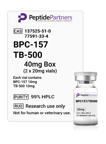
Combo Vial: BPC-157/TB-500 (20mg vials)
Price range: $224.00 through $880.00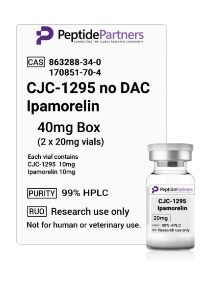
Combo Vial: CJC-1295 no DAC/Ipamorelin (20mg vials)
Price range: $220.00 through $877.00
GLOW Blend: BPC-157/TB-500/GHK-Cu (70mg vials)
Price range: $182.00 through $770.00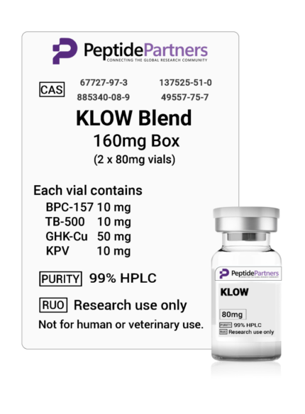
KLOW Blend: BPC-157/TB-500/GHK-Cu/KPV (80mg vials)
Price range: $240.00 through $975.00
